October is the month when your neighbour puts a skeleton on the porch and you quietly notice that its joints would benefit from a second opinion.
For Canadian healthcare practitioners, Halloween has a certain professional familiarity. Anatomical models become decorations. White coats become costumes. Someone buys a plastic stethoscope that looks unlikely to survive even a moderately enthusiastic cardiovascular examination.
Yet medicine’s own history contains objects and stories that need very little seasonal embellishment.
There are masks with beaks, wax figures with removable organs and photographs that made the invisible interior of the body suddenly visible. There is also an important Canadian medical story that begins on 31 October, in a bedroom in London, Ontario.
This Halloween, take a short tour through strange medical history, where the real explanations are often more interesting than the spooky versions.
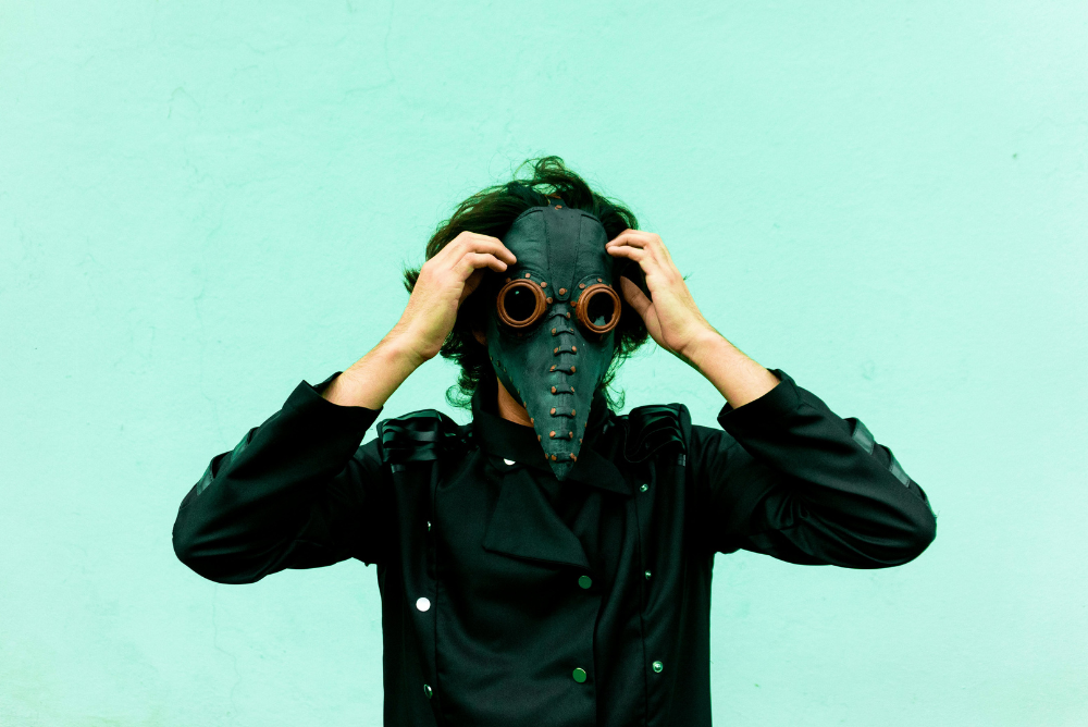
Few medical images are more recognisable at Halloween than the long-beaked plague mask.
It looks like something designed by a committee consisting of a physician, a crow and a very dramatic milliner.
The familiar explanation is that the beak held aromatic substances intended to protect the wearer from disease-causing air. Wellcome Collection’s account of plague clothing describes this connection to miasma theory, which associated infection with polluted or foul-smelling air.
However, the costume’s history is less straightforward than the Halloween aisle suggests.
A historical study of the plague doctor’s mask cautions against treating it as the standard uniform of medieval plague medicine. The authors place documented references to the beaked mask later, in the seventeenth century, particularly in Italy and southern France.
The famous image is not a reliable shortcut for several centuries of medical practice.
That distinction gives practitioners an excellent piece of seasonal trivia. The next time someone describes the costume as what every doctor wore during the Black Death, you have a more interesting story ready.
Whether the Halloween party wants a discussion of historical evidence is another matter. Read the room before opening the references.

A stethoscope is such a familiar professional object that its original design can feel unexpectedly improvised.
According to the Science Museum Group’s account of Laennec’s stethoscope, René Laennec used a tube of rolled paper in 1816 to listen to a young woman’s heart, avoiding the direct placement of his ear against her chest. He subsequently developed a wooden instrument.
The early device looks rather different from the equipment now draped around shoulders or temporarily misplaced in examination rooms.
One of medicine’s most recognisable instruments began with a practical response to an awkward encounter.
There is something appealingly human about that origin. Medical invention can sound grand in retrospect, complete with portraits and dates. At the beginning, however, there may be someone looking at an immediate problem and wondering whether a simple object could help.
Imagine presenting the prototype at a modern procurement meeting.
Does it connect wirelessly? No.
Does it require charging? Also no.
What happens if someone spills coffee on it? We should probably move on to the wooden version.
The story also makes the stethoscope a particularly good Halloween talking point. A costume accessory that appears ordinary has a history more surprising than its plastic packaging suggests.
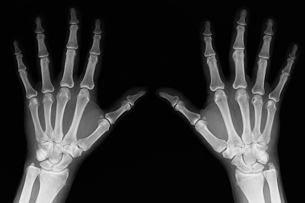
A skeletal hand is now an entirely predictable Halloween image.
In the late nineteenth century, seeing the bones inside a living person’s hand was a rather different proposition.
Wilhelm Conrad Röntgen discovered X-rays in 1895. Among the famous early images is his wife Anna Bertha’s hand, with its bones and ring visible. The image appears in the Nobel Prize organisation’s Röntgen photograph collection.
The Nobel account of the discovery’s public reception describes how rapidly the news attracted attention.
For a practitioner accustomed to reviewing imaging, it takes a little imagination to recover the strangeness of that moment.
The body’s interior could become an image without first being exposed by dissection or surgery.
Set aside the familiar reporting systems and viewing software for a moment. Think instead of encountering that possibility for the first time.
It is a reminder that something can become routine in clinical work while remaining extraordinary in principle.
The Halloween skeleton on the reception desk may have questionable proportions. The historical image of a living hand deserves a little more respect.
It also offers a useful challenge for anyone planning a medical trivia quiz: ask participants what made the image remarkable, rather than simply asking them to name its creator.
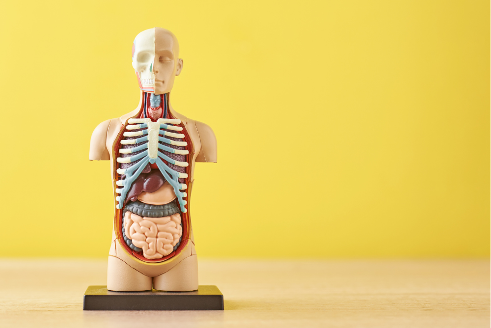
Before opening this particular historical cabinet, imagine an anatomical teaching model made with the visual care of a portrait.
Now imagine that some of its internal structures can be removed.
The Science Museum’s discussion of anatomical wax models includes a model by Francesco Calenzuoli with a removable heart. Such objects bring craftsmanship and anatomical representation into the same display case.
To a modern viewer, they can appear unsettling. To someone interested in medical education, they are also absorbing examples of how the body has been represented for study.
Context changes what an object seems to be.
Placed in a dimly lit Halloween display, a wax anatomical figure might suggest that the evening has taken an unfortunate turn. Presented with its history and educational purpose, it invites closer attention to the skill involved in making anatomy visible.
Practitioners already understand this change in perspective. An object that seems unusual to a visitor can be entirely ordinary in a teaching environment.
There is also a familiar practical question hiding behind any model with removable components: where does everything go when the session is finished?
Anyone who has encountered an anatomical model with an unexplained spare piece will appreciate that some educational challenges are timeless.

The strongest Canadian connection in this collection does not involve a mask or a skeleton.
It involves an idea.
In the early hours of 31 October 1920, Frederick Banting recorded a hypothesis while living in London, Ontario. Banting House National Historic Site describes the moment and its relationship to the work that later led to insulin’s discovery.
The date gives Canadian practitioners a Halloween fact with considerably more substance than another joke about frightening appointment schedules.
It is important, though, to distinguish the initial idea from the subsequent research.
Insulin was not discovered in a completed experiment that Halloween night. The later achievement involved the work of Banting, Charles Best, James Collip and John Macleod in Toronto. Parks Canada’s history of insulin’s discovery describes the collaborative effort and its significance.
That makes the story richer.
The memorable moment belongs in a bedroom in Ontario. The development that followed belongs to a larger account of investigation, collaboration and experimental work.
For an October staff-room quiz, try asking: “Which major Canadian medical discovery has an origin story connected to Halloween?”
It is an excellent question precisely because the answer is unlikely to be anyone’s first guess.
These histories can make an enjoyable seasonal feature for a practice newsletter or an optional staff-room quiz.
Keep it simple. Choose an object, ask a question and provide the explanation with a link to the source.
For example:
If sharing historical material with patients, choose images and wording appropriate to your audience. A curious story can work perfectly well without graphic pictures.
The appeal is in the surprise: an ordinary instrument with an unlikely beginning, a familiar costume with a misunderstood history, or a Canadian anniversary hiding in plain sight.
Medical practitioners spend much of their time using tools, concepts and techniques whose histories sit quietly in the background.
Halloween offers an excuse to bring a few of those stories forward.
The stethoscope has an inventive beginning. The skeletal hand has a place in the history of imaging. The beaked mask deserves a more careful explanation. And 31 October has a remarkable connection to Canadian medicine.
There is plenty here to keep a curious colleague entertained.
As for the porch skeleton’s anatomically ambitious knees, you may have to let those go.
It is only working one month of the year.
This article is for informational purposes and reflects developments as of July 2026. Consult official provincial sources for the latest policy updates.
Ready to start practicing better? Let’s talk! Get your questions answered or book a demo with GoodX Healthcare.